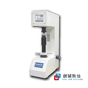
HTR-RAN高溫洛氏硬度計
HTR-RAN高溫洛氏硬度計1.產品概述該設備用來測定試樣從常溫到高溫環境下的維氏硬度值,用來測試金···
全自動橡膠硬度計
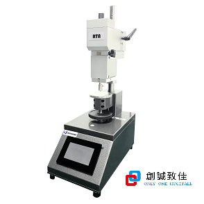
全自動橡膠硬度計RTA多種尺規可組裝Shore: A, A0, 0, C, D, D0, 00, 0···
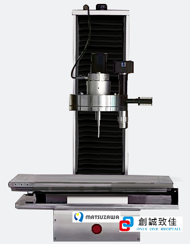
全自動洛氏硬度計
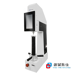
日本Matsuzawa RANS自動全洛氏硬度計,該原裝進口硬度計為電子閉環加載,全自動測量,可測量···
動態文章
二氧化碳培养箱是通过在培养箱箱体内模拟形成一个类似细胞/组织在生物体内的生长环境,培养箱要求稳定的温度(37°C)、稳定的CO2水平(5%)···
Posted in 技術文章 by Admin on 2023-10-12 16:50:15
拋光是通過摩擦或應用化學處理來創建光滑且有光澤的表面的過程,留下具有顯著鏡面反射的幹淨表面。在某些材料(例如金屬、玻璃、黑色或透明石頭)中,···
Posted in 技術文章 by Admin on 2023-10-10 10:03:32
硬度壓頭是硬度計上壓入試件而無**變形的零部件。常用的硬度壓頭有洛氏硬度壓頭、布氏硬度壓頭、維氏硬度壓頭、努氏硬度壓頭以及一些硬度計壓頭等。
Posted in 技術文章 by Admin on 2023-10-09 17:14:07
奧林巴斯創立於1919年,至今已經有一百多年。奧林巴斯是一家精密光學與成像的日本公司,以顯微鏡事業起家。顯微鏡的透鏡研磨技術被譽為“獨門秘笈···
Posted in 技術文章 by Admin on 2023-10-08 11:40:31
創誠致佳
創誠致佳公司是中國大陸專業從事硬度計等儀器設備銷售,公司具有非常豐富的硬度計現場應用經驗,可定制各種應用場景的硬度測試設備等,客戶有設備測試應用場景,創誠致佳可以根據客戶需求來實現,這得益於公司豐富的行業應用經驗。讓客戶愛上我們的每一套設備是我們的願景。